Salvia microphylla 에탄올추출물의 항산화 및 항염에 관한 연구
A Study on Antioxidant and Anti-inflammatory of Salvia microphylla Ethanol Extract
樱桃鼠尾草(Salvia microphylla)乙醇提取物的抗氧化以及抗炎研究
Article information
Abstract
목적
본 연구는 Salvia microphylla 에탄올추출물(SME)의 항산화 활성 및 항염 활성을 확인하는 목적으로 진행되었다.
방법
본 연구에서 SME은 50% 에탄올을 용매로 상온에서 7일간 정치하여 추출하였다. 항산화 활성은 총 폴리페놀, 플라보노이드, DPPH 및 ABTS radical 소거능 방법을 사용하여 측정하였다. 항염 활성은 LPS 처리된 RAW 264.7 세포로부터 NO 생성 감소와 iNOS 단백질 발현량 감소를 확인하여 검증하였다.
결과
본 연구에서는 SME의 총 폴리페놀과 총 플라보노이드 함량은 102.26±0.39 mg GAE/g, 377.42±0.77 mg QUE/g를 나타내었다. DPPH와 ABTS는 50-1000 μg/mL에서 농도의존적 항산화 활성을 나타내었다. SME의 세포생존율은 50-1000 μg/mL에서 99% 이상으로 세포독성은 없는 것으로 나타내었다. SME은 농도의존적으로 NO 생성을 감소 효과를 나타내었으며, 600 μg/mL에서 0.00±1.34%로 100%에 가까운 NO 생성 억제 효과를 확인하였다. 더욱이 염증관련 인자인 iNOS의 단백질 발현량은 농도의존적인 감소 효과로 항염 활성을 확인하였다. SME이 코스메슈티컬의 성분으로 잠재력을 확인할 수 있었다.
결론
본 연구는 이와 같은 결과들로부터 SME은 화장품에서 항산화 및 항염 활성이 있는 천연방부제 및 피부 자극 완화와 관련된 원료로서 유용하게 활용 가능할 것으로 사료된다.
Trans Abstract
Purpose
This study was aimed at investigating the anti-inflammatory and antioxidant properties of <ji>Salvia microphylla (SM) extract (SME).
Methods
Powdered SM was treated with ethanol and decocted to acquire SME. The antioxidant activities of SME were assessed by evaluating its total polyphenolic, and flavonoid , contents and performing 2,2-diphenyl-1-picryl-hydrazyl-hydrate (DPPH) and 2,2′-azino-bis(3-ethylbenzothiazoline-6-sulfonic acid) (ABTS) radical scavenging assays. The anti-inflammatory activities of SME were verified by examining the inhibition of nitric oxide (NO) production, and inducible nitric oxide synthase (iNOS) expression in lipopolysaccharide (LPS) activated RAW 264.7 macrophages.
Results
The total polyphenol and flavonoid contents in SME were 102.26±0.39 mg GAE/g and 377.42±0.77 mg QUE/g, respectively. The DPPH and ABTS assays revealed the antioxidant activities of SME by exhibiting their ability to scavenge radicals in a concentration-dependent manner (50-1000 μg/mL). The cell viability of SME exceeded 99% at 50-1000 μg/mL. The concentration-dependent reduction in the expression of the inflammation mediator iNOS and subsequent NO production indicated an approximately 100% inhibition efficacy at 600 μg/mL (0.00±1.34%). These results confirmed the potential of SME as a component of cosmeceuticals.
Conclusion
Based on the findings of this study, SME is explicitly acknowledged for its applicability as a natural preservative possessing antioxidant and anti-inflammatory activities as well as a raw material for cosmeceuticals intended to alleviate skin irritation.
Trans Abstract
目的
本研究旨在研究樱桃鼠尾草 (Salvia microphylla,SM) 提取物 (SME) 的抗炎和抗氧化特性。
方法
将SM粉末用乙醇处理,煎煮得SME。通过评估其总多酚和黄酮类化合物的含量,并测定DPPH和ABTS自由基清除活性来评估SME的抗氧化活性。通过检测脂多糖 (LPS) 激活的RAW 264.7巨噬细胞中一氧化氮 (NO) 产生和诱导型一氧化氮合酶 (iNOS) 表达的抑制作用,验证了SME的抗炎活性。
结果
SME中总多酚和黄酮含量分别为102.26±0.39 mg GAE/g和377.42±0.77 mg QUE/g。DPPH和ABTS检测通过显示SME以浓度依赖性方式 (50-1000 μg/mL) 清除自由基的能力,揭示了SME的抗氧化活性。SME在50-1000 μg/mL时,细胞活力超过99%。炎症介质iNOS表达和随后的NO产生呈浓度依赖性减少,表明600 μg/mL时的抑制功效约为100%(0.00±1.34%)。这些结果证实了SME作为药妆品成分的潜力。
结论
根据本研究的结果,SME因其作为具有抗氧化和抗炎活性的天然防腐剂以及旨在减轻皮肤刺激的药妆原料的适用性而得到明确认可。
Introduction
현대를 살고 있는 인류는 생활환경과 식습관의 변화로 생활습관병이 증가하고 생물학적, 화학적 유해 환경 등에 빈번히 노출되어 생체 내의 면역 조절 기능 이상으로 알러지, 아토피 등의 염증성 피부질환이 증가하고 있다(Kim et al., 2022). 화학적으로 합성된 항염제제의 장기간 사용에 따른 부작용 사례들이 보고되면서 천연소재를 활용한 항염 활성 물질 탐색이 진행되고 있으며, 또한 항산화능을 가진 천연소재들은 페놀산과 플라보노이드 등을 함유하고 있으며, 이를 많이 함유하고 있는 천연소재들은 뛰어난 항염 효과가 보고된 결과들을 바탕으로 항산화 활성이 우수한 천연물들의 항염 활성을 확인하는 연구들이 활발히 수행되고 있다(Kim et al., 2017; Namiki, 1990). 염증반응은 다양한 유형의 감염으로 발생하거나 대사물질 또는 외부의 물리적, 화학적 자극에 의한 손상으로부터 생체를 보호하기 위한 생체조직의 방어 기전으로 신체 손상을 회복하는 반응이다(Lee & Kim, 2018; Shim, 2018; Kim et al., 2022). 염증반응에 관여하는 세포인 macrophage, monocyte, 비만세포 등은 활성산소종(reactive oxygen species, ROS)과 활성질소종(reactive nitrogen species), prostagrandin E2 (PGE2), 염증성 cytokine 등의 염증 매개체들을 분비한다(Lee & Kim, 2018). 세포의 nitric oxide synthase (NOS)에는 neuronal NOS (nNOS), endothelial NOS (eNOS), inducible NOS (iNOS)이 있으며 이중 iNOS가 염증반응에 주로 관여하며, interferon-γ (IFN-γ), liopopolysaccharide (LPS), 염증성 사이토카인의 자극이 있을 때 발현된다. 염증 시 iNOS에 의해 생성된 NO는 염증반응을 촉진시킨다(Jung & Cho, 2011; Kim et al., 2022). 따라서 NO의 생성이나 PGE2, ROS, 염증성 cytokine 등의 발현억제는 염증성 질환 치료 예방에 주요한 역할을 한다(Lee & Kim, 2018; Shim, 2018).
Salvia microphylla는 멕시코가 원산지인 꿀풀과(Lamiaceae) Salvia속에 속하는 식물이며 "Hotlip sage" 라고도 불린다(Fair et al., 2012). Salvia microphylla 차는 전통적으로 복통과 설사와 같은 위장 문제를 치료하고 불면증 치료에 사용되어왔다(Jenks & Kim, 2013; Pérez-Nicolás et al., 2018). 부인과 질병에 사용되었으며(Jenks & Kim, 2013), 항산화(Cha et al., 2009), 항종양제(Mathew & Thoppil, 2012) 항박테리아, 리파아제 억제제, 유리라디칼 억제제(Villa-Ruano et al., 2013), 살충효과(Zavala-Sánchez et al., 2013; Romo-Asunción et al., 2016) 등에 효과가 있다고 보고되었다. 그러나 SME에 대한 RAW 264.7 cell의 염증 완화에 관련된 연구가 미비한 실정이다. 따라서 본 연구에서는 LPS에 의해 자극이 유발된 RAW 264.7 cell에 SME의 항산화 활성과 항염 효과를 확인하여 기능성 화장품 소재로서의 활용 가능성을 확인하고자 하였다.
Methods
1. 추출물 제조
본 실험에서 분석을 위해 사용된 S. microphylla는 경상남도 거창군 가조면에 소재하는 플라워쉐프 농장에서 채취하였다. 추출물은 S. microphylla 파우더 20 g과 50% 에탄올 400 mL을 용매로 상온에서 7일간 정치하여 추출하였으며, 추출물은 여과지(Whatman No.2; GE Healthcare, UK)를 사용하여 2회 여과하고, 감압 하에서 Rotary Evaporator (Eva-5; DAIHAN Scientific, Korea)로 농축하고, 용매를 제거한 후 본 실험에 사용할 때까지 냉동 보관하였다.
2. 세포 배양
본 실험에 사용된 마우스 대식세포 계열 RAW 264.7 cell (murine macrophage cell line) 은 American Type Culture Collection (ATCC, USA)에서 분양 받아 사용하였다. RAW 264.7 cell는 10% fetal bovine serum (FBS; Gibco BRL, USA)와 1% penicillin/streptomycin (Gibco BRL)을 넣은 Dulbecco's Modified Essential Medium (DMEM; HyClone, Cytiva, USA)에서 배양하였다. 세포는 37℃, 5% CO2 세포배양기에서 배양하였다.
3. 총 폴리페놀 함량 측정
총 폴리페놀 함량은 Folin-Ciocalteu 방법을 일부 변형하여 분석하였다(Ainsworth & Gillespie, 2007). 총 polyphenol을 측정하기 위해 gallic acid (Daejung chemicals & metals Co., LTD., Korea)를 농도별(0-800 μg/mL)로 희석하여 사용하였다. SME 또는 gallic acid 50 μL와 50% Folin-Ciocalteu reagent (Sigma-Aldrich, USA) 50 μL를 혼합 후 3 min 반응하였다. 그 후 2% Na2CO3 1 mL과 혼합 후 30 min 반응시키고, 700 nm에서 microplate reader (Molecular Devices, USA)를 이용하여 흡광도를 측정하였다. 총 polyphenol 함량은 건조추출물 무게에 대한 gallic acid의 표준곡선과 비교하여 mg GAE/g로 표현하였다.
4. 총 플라보노이드 함량 측정
총 플라보노이드 함량은 aluminum chloride colorimetric 방법을 일부 변형하여 분석하였다(Zhishen et al., 1999). 총 flavonoid를 측정하기 위해 quercetin (Sigma-Aldrich)을 농도별(0-1 mg/mL)로 희석하여 사용하였다. SME 또는 quercetin 25 μL, 증류수 125 μL, 5% NaNO2 10 μL를 혼합 후 5 min 반응하였다. 반응 후 10% AlCl3 15 μL를 첨가하고 6 min 반응시켰다. 그 후 1 M NaOH 50 μL를 첨가한 후, 510 nm에서 microplate reader를 이용하여 흡광도를 측정한다. 총 flavonoid 함량은 건조 추출물 무게에 대한 quercetin의 표준곡선과 비교하여 mg QUE/g로 표현하였다.
5. DPPH radical 소거능 측정
항산화능 분석을 위하여 2,2-diphenyl-1-picrylhydrazyl (DPPH; Alfa Aesar, USA) radical을 일부 변형하여 측정하였다(Blois, 1958). 0.2 mM의 DPPH용액 60 μL에 농도별(50-1000 μg/mL)로 희석한 시료 120 μL를 96 well plate에 처리하였다. 암실에서 15 min 반응시킨 후 microplate reader를 이용하여 517 nm에서 흡광도를 측정하였다.
6. ABTS radical 소거능 측정
ABTS radical 소거활성 측정은 potassium persulfate와의 반응에 의해 생성되는 ABTS free radical이 항산화제에 의해 소거되어 청색이 사라지는 것을 이용하여 측정하였다(Re et al., 1999). 7 mM 2,2'-azinobis(3-ethylbenzthiazoline-6-sulfonic acid (ABTS; WAKO, Japan) 시약 100 μL에 농도별(50-1000 μg/mL)로 희석한 시험물질 50 μL를 96-well plate에 처리하였다. 암실에서 5 min 반응시킨 뒤 microplate reader로 734 nm에서 흡광도를 측정하였다.
7. 세포 생존율 측정
세포 생존율은 MTT (3-(4,5-Dimethyl-2-thiazolyl)-2,5-diphenyl-2H-tetrazolium bromide) assay를 통해 확인하였다. RAW 264.7 cell를 96 well plate (3×10³ cells/well)에 분주한 다음, 16-24 h 동안 배양하였다. 그 후 추출물을 50-1000 μg/mL로 처리한 다음 37℃, 5% CO2 조건에서 24 h 배양하였다. 세포의 생존율을 확인하기 위해서 well 당 MTT (Sigma-Aldrich) (5 mg/mL)를 10 μL 처리하여 37℃, 5% CO2 배양기에서 30 min 반응시킨 후 상등액을 제거하였다. 형성된 formazan을 dimethyl sulfoxide (DMSO; Sigma-Aldrich) 150 μL로 녹이고 microplate reader를 이용하여 540 nm에서 흡광도를 측정하였다.
8. NO 생성 억제능 측정
NO 생성 억제율을 확인하기 위하여 24 well plate에 RAW 264.7 cell를 2.5×10⁵ cells/well로 분주하고 37℃, 5% CO2 incubator 조건하에 24 h 배양한 후 실험에 사용하였다. 배양시킨 cell을 1 μg/mL LPS (Sigma-Aldrich)가 포함된 배지로 교환 후, SME을 농도별(50-1000 μg/mL)로 첨가하여 24 h 배양하였다. 배양 후 상층액을 얻어, 세포배양액 중으로 분비한 NO 양을 griess reagent system (Promega, USA)를 이용하여 측정하였다. 생성된 NO 양은 sodium nitrite (NaNO2)를 이용하여 검정곡선을 작성하여 비교하였다
9. Western blot 분석을 통한 단백질 발현 측정
항염 효과를 확인하기 위해 RAW 264.7 cell (1×10⁶ cells)을 100 mm 배양접시에 분주하여 37℃, 5% CO2 incubator 조건하에 24 h 동안 배양 후, 상등액 제거, phosphate buffer saline (PBS)로 세척 후, SME를 농도별로 1 h 처리한 뒤 LPS 1 μg/mL 처리 후에 24 h 추가 배양하였다. 배양 후 Radioimmunoprecipitation assay buffer (RIPA buffer; Cell signaling, USA)를 이용하여 용해한 후, 원심분리기(Centrifuge 5430R; Eppendorf, Germany)를 이용하여 원심분리(10,000×g, 10 min)로 단백질을 분리하였으며, BCA protein assay kit (iNtron Biotechnology, Inc., Korea)로 단백질을 정량하였다. 12% polyacrylamide SDS gel에서 전기영동 한 후, ㅔ olyvinylidene fluoride (PVDF; Sigma-Aldrich) membrane으로 이동시켰다. PVDF membrane을 blocking buffer (3% skim milk; BD Difco, USA) 로 2 h blocking하고 primary antibody (CST, USA)를 blocking buffer에 희석하여 4℃에서 overnight 하였다. PBS-T (0.05% Tween 20 in PBS)로 washing 후 secondary antibody (CST, USA)를 blocking buffer에 희석하여 실온에서 반응시켰다. PBS-T로 washing하고 West Pico PLUS Chemiluminescent Substrate (Thermo Scientific, USA)에 반응 후, Chemiluminescent imaging system (Fusion Solo S, VILBER, France)을 이용하여 단백질 발현을 측정하였다. Image J program (National Institutes of Health, USA)을 사용하여 상대적인 단백질 발현을 정량하였다.
10. 통계처리
실험결과는 mean±SD로 표기하였으며, Graphpad Prism software (version 5.00 for Windows, USA) 프로그램을 이용한 one way ANOVA를 실시한 후 Tukey's test 방법을 사용하여 그 결과 p<0.05 수준에서 유의성을 검증하였다.
Results and Discussion
1. S. microphylla 에탄올추출물의 항산화 효과
가. 총 폴리페놀과 플라보노이드 함량 평가
페놀 화합물은 식물계에 널리 분포되어 있으며, 다양한 구조와 분자량을 가지고 있는 2차 대사산물로서 phenolic hydroxy기를 가지고 있어 단백질과 같은 큰 분자들과 결합하는 성질이 있으며, 항산화의 생리활성 기능을 가지고 있다(Park & Ryu, 2022).
플라보노이드는 식물의 꽃, 줄기 및 열매 등에 많이 함유되어 있으며, 항산화, 항염 효과, 면역개선, 항바이러스 등 여러 생리활성 작용이 있다(Park & Na, 2020). 세이지 종들은 페놀 화합물에 기반한 항산화 특성으로 널리 사용되고 있다(Poulios et al., 2020).
SME의 총 폴리페놀와 플라보노이드의 함량은 각각 102.26±0.39 mg GAE/g과 377.42±0.77 mg QUE/g으로 확인되었다(Table 1). 플라보노이드는 ROS를 제거하여 항산화 효능이 우수하다고 보고되어졌으며, 폴리페놀과 같이 항바이러스, 항염증, 항암 효과가 보고되어졌다(Kim et al., 2012).

Total phenolic and flavonoid contents of Salvia microphylla ethanol extract
나. DPPH radical 소거능 평가
SME의 DPPH radical 소거능을 측정한 전자공여능(EDA) 결과를 Figure 1에 나타내었다. DPPH는 radical 소거능을 평가하기 위하여 일반적으로 사용된다. DPPH radical 소거능은 보라색의 DPPH가 항산화 물질에 의해 환원되면서 노란색으로 탈색되는 원리를 이용하여 산화 방지제 활성을 측정하며 그 측정 방법이 비교적 간단하여 여러 시료로부터 항산화 활성을 탐색할 때 유용하다(Huang et al., 2005). SME는 50-1000 μg/mL에서 농도의존적으로 소거활성을 나타내었다. 200 μg/mL에서 79.00±0.16%, 400 μg/mL에서 85.91±0.17%, 600 μg/mL에서 86.12±1.00% 및 1000 μg/mL에서 88.20 ±0.39%의 소거활성을 나타내었다. 세이지 종인 Salvia officinalis L 60% 에탄올 추출물은 200 μg/mL에서 66.3±2.2%의 DPPH radical 소거능이 보고되었다(Cha et al., 2009). 따라서 SME의 DPPH radical 소거능이 우수하다고 판단할 수 있었다.
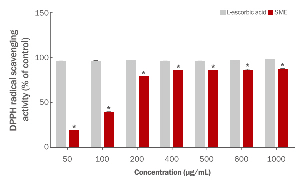
DPPH radical scavenging activity of Salvia microphylla ethanol extract.
DPPH radical scavenging assays were conducted to investigate the antioxidant effects of SME at various concentration. The values are then expressed as mean±SD of three independent experiments (*p<0.05). SME, Salvia microphylla ethanol extract.
다. ABTS radical 소거능 평가
ABTS radical 소거능은 항산화 측정방법으로 단기간에 측정이 가능하며, 시료의 항산화제가 potassium persulfate과 반응하여 생성된 ABTS radical을 제거한다는 점을 이용하여 지용성과 수용성 물질을 모두 측정할 수 있다(Um & Ryu, 2022).
Cha et al. (2009) 연구에서는 세이지 종인 Salvia officinalis L 60% 에탄올 추출물은 200 μg/mL에서 97.6±0.1%의 ABTS radical 소거능이 보고되었다. SME의 ABTS radical 소거능은 50-1000 μg/mL에서 농도의존적으로 소거활성을 나타내었다. 400 μg/mL에서 80.87±0.54%, 600 μg/mL에서 94.92±0.87% 및 1000 μg/mL에서 99.27±0.13%의 소거활성을 나타내었다(Figure 2). 이와 같이 SME는 농도의존적으로 ABTS radical 소거능이 우수하다고 판단할 수 있었다.
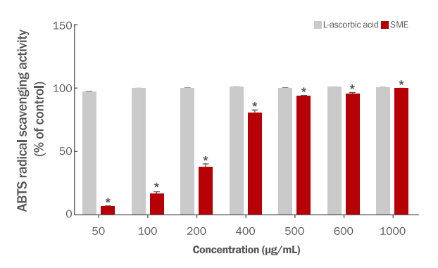
ABTS radical scavenging activity of Salvia microphylla ethanol extract.
ABTS radical scavenging assays were conducted to investigate the antioxidant effects of SME at various concentration. The values are then expressed as mean±SD of three independent experiments (*p<0.05). SME, Salvia microphylla ethanol extract.
2. 세포 생존율
SME가 대식세포인 RAW 264.7 cell에 미치는 생존율을 측정하기 위하여 MTT assay를 이용하였다. SME를 50-1000 μg/mL 농도로 처리하여 24 h 배양시킨 후 대조군과 비교하였다. SME는 50 μg/mL에서 101.03±5.12%, 200 μg/mL에서 122.04±4.89%, 400 μg/mL에서 134.20±3.88% 및 1000 μg/mL에서 99.09±6.51%의 세포생존율을 나타내었다(Figure 3). 세포 생존율은 50-1000 μg/mL의 전체 농도에서 99% 이상으로 ISO 10993-5 기준에 의거하여 본 실험에 사용된 SME의 실험농도에서는 세포독성이 나타내지 않은 것으로 확인되었다. 따라서 이후 실험은 99% 이상의 생존율을 나타내는 50-1000 μg/mL 농도에서 진행하였다.
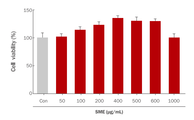
Effect of Salvia microphylla ethanol extract on cell viability of RAW 264.7 cell.
RAW 264.7 cells were treated with various concentration of SME (50–1000 μg/mL) using MTT assay. The responses in the control in each test are expressed as 100%. Each value expresses the mean±SD of three independent experiments. Con, none treated control; SME, Salvia microphylla ethanol extract.
3. NO 생성 억제능 평가
NO의 생성이나 PGE2, ROS, 염증성 cytokine 등의 발현억제는 염증성 질환을 치료하거나 노화 예방에 주요한 역할을 한다(Lee & Kim, 2018; Shim, 2018). NO는 조직과 신경 손상을 유발하고, 유전자 변이를 유도하거나, 부종을 유발하는 등 과도한 염증반응을 일으킨다(Shim, 2018).
LPS만 처리한 대조군의 NO 생성량을 기준(100%)으로 SME의 NO 생성은 50-1000 μg/mL에서 농도의존적으로 감소되는 것을 확인하였다. 600 μg/mL에서는 0.00±1.34%로 100%에 가까운 억제효과를 나타내어 SME의 함염 활성이 우수한 것으로 판단할 수 있었다(Figure 4).
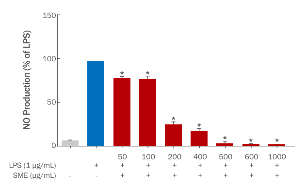
Effect of Salvia microphylla ethanol extract on nitric oxide (NO) production in RAW 264.7 cell.
RAW 264.7 cells were treated with SME and LPS (1 μg/mL). Each value expresses the mean±SD of three independent experiments (*p<0.05). LPS, lipopolysaccharide treated control; SME, Salvia microphylla ethanol extract.
4. Western blot 분석을 통한 iNOS 단백질 발현억제 효과
염증 관련 인자인 NOS는 3가지의 isoform으로 NOS I (neuronal NOS, nNOS), NOS II (inducible NOS, iNOS), NOS III (endothelial NOS, eNOS)가 알려져 있으며, nNOS와 eNOS는 항상 발현되어 있어서 constitutive NOS (cNOS)라하며, iNOS는 대식세포 등에서 cytokine, LPS와 같은 내독소의 자극에 의해 발현되어 NO 생성을 촉진시켜 세포독성, 염증성 질환 등의 작용이 발생한다(Yim, 2010; Cossenza et al., 2014). 이러한 염증 관련 인자에 대한 억제효과를 확인하여 항염 활성을 확인하고자 하였다.
SME에 대해 iNOS의 단백질 발현은 Western blot을 통해 측정한 결과를 Figure 5에 나타내었다. iNOS의 단백질 발현량은 100 μg/mL (74.84±5.95%), 200 μg/mL (74.68±3.53%), 400 μg/mL (60.43±2.87%), 600 μg/mL (50.72±7.64%) 및 1000 μg/mL (10.08±5.78%)로 농도의존적으로 감소되는 것을 확인하였다. 결과와 같이 SME는 NO 소거능과 iNOS 발현억제에 의한 항염 효과가 있을 것으로 판단되었다.
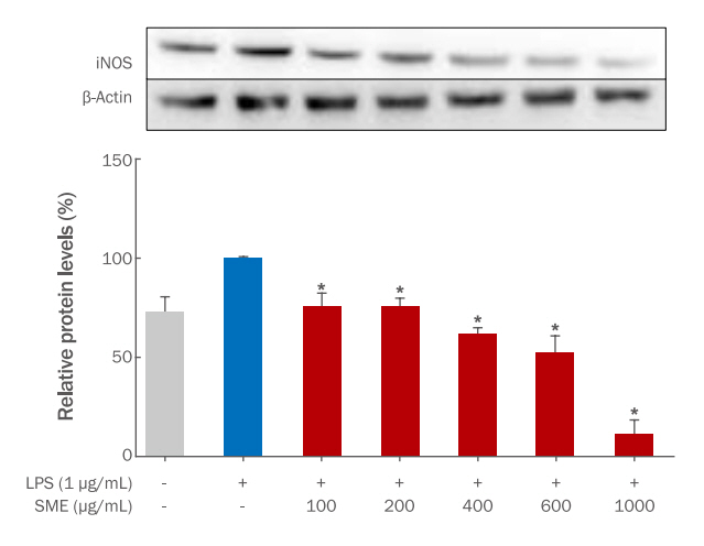
Effect of Salvia microphylla ethanol extract on iNOS protein expression in RAW264.7 cells.
RAW 264.7 cells were pretreated SME at various concentration (100-1000 μg/mL) for 1 h and then treated LPS (1 μg/mL) for 24 h. iNOS protein expression was determined in RAW 264.7 cells treated with SME. Each value expresses the mean±SD of three independent experiments (*p<0.05). LPS, lipopolysaccharide treated control; SME, Salvia microphylla ethanol extract.
세이지 종에는 가장 중요한 생물학적 활성 화합물로 quercetin, rosmarinic acid, caffeic acid, linalool, luteolin-7-glucoside, camphor, 1,8-cieole, α-pinene이다(Poulios et al., 2020). 이 중 caffeic acid 및 유도체는 항염 작용으로 NO 소거능뿐만 아니라, iNOS 발현 억제가 보고되었다(Da Cunha et al., 2004).
Conclusion
최근 현대 사회는 도시화와 산업의 발전으로 유해 환경으로부터의 수 많은 노출과 식생활 변화 등에 의한 생체 면역 조절 이상으로 알러지, 아토피 및 염증 등의 질환이 증가하고 있다(Kim et al., 2022).
본 연구에서는 SME의 항산화 활성 및 항염 활성 등을 평가하여 SME의 피부질환 개선 기능성 소재로서 이용 가능성을 확인하고자 하였다. 항산화 활성은 폴리페놀 함량, 플라보노이드 함량, DPPH 및 ABTS를 이용하여 확인하였다. 그 결과, SME의 총 폴리페놀과 플라보노이드는 각각 102.26±0.39 mg GAE/g과 377.42±0.77 mg QUE/g, DPPH radical 소거활성은 농도의존적인 활성을 나타내었으며, 200 μg/mL에서 75% 이상의 소거능을 확인하였다. 또한 ABTS radical 소거활성도 농도의존적인 반응을 나타내었으며, 400 μg/mL에서 80% 이상의 소거능을 확인하였다. 전체적인 농도로 보았을 때 SME의 항산화 활성이 뛰어난 것으로 확인되었다. 세포 생존율은 50-1000 μg/mL에서 99% 이상의 세포 생존율을 나타내어 SME의 세포 독성이 없음을 확인하였다. 항염 활성은 SME의 NO 생성억제와 iNOS 단백질 발현억제 효과를 확인하여 항염 활성을 확인하였다. SME의 NO 생성억제 활성은 50-1000 μg/mL에서 농도의존적인 감소를 나타내었고, 600 μg/mL에서 0.00±1.34%로 100%에 가까운 NO 생성억제효과를 확인하였다. 염증 관련 인자인 iNOS 단백질 발현량도 600 μg/mL에서 50.72±7.64%, 1000 μg/mL에서 10.08±5.78%로 감소하는 것을 확인하였다. 이와 같은 결과로부터 SME는 NO 소거능뿐만 아니라 iNOS 발현억제에 의한 염증 억제능이 있는 것으로 판단되어 S. microphylla 에탄올추출물이 화장품에서 항산화 및 항염 활성이 있는 기능성화장품 원료로서 유용하게 활용 가능할 것으로 사료된다.
Acknowledgements
이 성과는 정부(과학기술정보통신부)의 재원으로 한국연구재단의 지원을 받아 수행된 연구임(No. 2021R1G1A1094165).
Notes
Author's contribution
IHC designed all the experimental investigations. Biological activity experiments were conducted together by GWC and HMJ, and IHC oversaw the project and contributed to all aspects of analysis and experimental design.
Author details
Guen Won Choi (Graduate Student), Department of Biomaterials Science, Gyeongsang National University, 33 Dongjin-ro, Jinju-si, Gyeongsangnam-do, 52725, Korea; Hyeon Mi Jo (Graduate Student), Department of Biomaterials Science, Gyeongsang National University, 33 Dongjin-ro, Jinju-si, Gyeongsangnam-do, 52725, Korea; In Ho Choi (Professor) Department of Plant and Biomaterials Science, Gyeongsang National University, 33 Dongjin-ro, Jinju-si, Gyeongsangnam-do, 52725, Korea.