Salvia microphylla 추출물의 항산화, 콜라겐 합성 및 MMP-1 저해활성 연구
A Study on Antioxidant, Pro-collagen Synthesis and MMP-1 Inhibitory Activities of Salvia microphylla Extract
小叶鼠尾草提取物的抗氧化、胶原蛋白合成及 MMP-1 抑制活性研究
Article information
Abstract
목적
본 연구는 S. microphylla 에탄올 추출물을 이용하여 항산화 활성, 콜라겐 합성 및 MMPs의 저해효과를 확인하는 목적으로 진행되었다.
방법
S. microphylla 추출물은 70% 에탄올을 용매로 사용하여 24 h 정치하여 추출하였다. S. microphylla 70% 에탄올 추출물의 항산화 효과를 확인하기 위하여 총 폴리페놀 함량, 총 플라보노이드 함량, DPPH radical 소거능 및 ABTS radical 소거능을 확인하였다. 섬유아세포(CCD-986sk)의 세포 생존율은 MTT 방법을 이용하였다. 섬유아세포에서 Type Ⅰ collagen 생성능과 MMP-1 단백질 발현 저해 활성을 통해 주름개선 효능을 확인하였다.
결과
실험에서 S. microphylla 70% 에탄올 추출물의 총 폴리페놀 함량은 198.22±2.14 mg GAE/g, 총 플라보노이드 함량은 291.55±6.60 mg QUE/g을 나타내었다. DPPH radical 소거활성은 400 μg/mL 농도에서 92.99±0.14%의 소거능을 나타내었고, ABTS radical 소거활성은 600 μg/mL 농도에서 99.37±0.12%의 소거능을 나타내었다. S. microphylla 70% 에탄올 추출물의 Type Ⅰ collagen 생합성은 농도의존적으로 증가하였으며, 200 μg/mL 농도에서 140.74±5.31%의 증가를 나타내었으며, MMP-1 생성은 농도의존적으로 감소하였으며, 200 μg/mL 농도에서 65.36±1.40%로 감소되었다. 따라서, S. microphylla 70% 에탄올 추출물은 주름 개선에 기여할 것으로 판단된다.
결론
이와 같은 결과로 S. microphylla 70% 에탄올 추출물은 우수한 항산화 활성과 pro-collagen 합성 증가와 MMP-1 생성의 저해 효과를 확인하여 기능성화장품의 기능성 소재로서 활용 가능성을 확인하였다.
Trans Abstract
Purpose
This study aimed to investigate the physiological activity of the antioxidant and anti-wrinkle effects of an ethanol extract of Salvia microphylla (SM) and the possibility of their application in new cosmetic material.
Methods
SME70 extract was prepared from powdered SM using 70% ethanol. For the analysis of the antioxidant activities of SME70, the total polyphenolic and flavonoid content, 2,2-diphenyl-1-picrylhydrazyl (DPPH), and 2,2′-azino-bis(3-ethylbenzothiazoline-6-sulfonic acid) (ABTS) radical scavenging assays were performed. The cell viability was determined in fibroblast cells (CCD-986sk) using an MTT assay. For analysis of the anti-wrinkle effects of SME70, pro-collagen synthesis and MMP-1 inhibition activities in human fibroblasts were investigated.
Results
The total polyphenol and flavonoid contents in SME70 were 198.22±2.14 mg GAE/g and 291.55±6.60 mg QUE/g, respectively. The DPPH radical scavenging activity of SME70 was 92.99±0.14% at 400 μg/mL and ABTS radical scavenging activity of SME70 was 99.37±0.12% at 600 μg/mL. The cell viability of fibroblasts was 99.56±2.77% at 200 μg/mL. Pro-collagen synthesis activity of SME70 increased in a concentration-dependent manner and was 140.74±5.31% at 200 μg/mL. The matrix metalloprotease (MMP-1) inhibition effect of SME70 decreased in a concentration-dependent manner, and was 65.36±1.40% at 200 μg/mL. Therefore, it is suggested that SME70 can be used as a new material for cosmetics with anti-wrinkle effects.
Conclusion
The antioxidant, pro-collagen synthesis, and MMP-1 inhibitory effects of SME70 were established, confirming its potential as a material for functional cosmetics with antiwrinkle properties.
Trans Abstract
目的
本研究旨在探讨小叶鼠尾草乙醇提取物的抗氧化和抗皱作用的生理活性及其在新型化妆品材料中应用的可能性。
方法
使用70%乙醇从SM粉末中制备SME70提取物。为了分析SME70的抗氧化活性,总多酚和类黄酮含量、2,2-二苯基-1-三硝基苯肼 (DPPH) 和 2,2'-连氮基-双(3-乙基苯并噻唑啉-6-磺酸) (进行了ABTS)自由基清除测定。使用MTT测定法测定成纤维细胞(CCD-986sk) 中的细胞活力。为了分析 SME70 的抗皱效果,研究了人成纤维细胞中的前胶原蛋白合成和MMP-1抑制活性。
结果
SME70中总多酚和黄酮含量分别为198.22±2.14 mg GAE/g和291.55±6.60 mg QUE/g。SME70在400μg/mL时的DPPH自由基清除活性为92.99±0.14%,SME70在600μg/mL时的ABTS自由基清除活性为99.37±0.12%。200 μg/mL时成纤维细胞的细胞活力为99.56±2.77%。SME70 的前胶原合成活性以浓度依赖性方式增加,在200 μg/mL时为140.74±5.31%。SME70对基质金属蛋白酶(MMP-1)的抑制作用呈浓度依赖性下降,在200 μg/mL时为65.36±1.40%。因此,建议SME70可作为具有抗皱功效的化妆品新原料。
结论
SME70的抗氧化、前胶原蛋白合成和 MMP-1 抑制作用得到证实,证实了其作为抗皱功能性化妆品材料的潜力。
Introduction
피부 노화는 연령 증가, 스트레스와 같은 내적 요인과 자외선(ultraviolet), 유해환경과 같은 외적 요인에 의해 영향을 받는 복잡한 생화학적 과정이다(Gragnani et al., 2014; Kim et al., 2013). 노화의 내적 요인인 연령 증가는 섬유아세포의 작용과 그 수의 감소로 인하여 탄력섬유의 변성과 교원섬유의 양 감소와 구조 변화로 탄력 저하를 발생시킨다(Kim et al., 2013; Kim et al., 2019). 진피층의 80-90% 존재하는 주요 구조 단백질인 Collagen (Type Ⅰ, Ⅱ, Ⅲ and Ⅳ)은 세포외 기질(extracellular matrix, ECM)의 구성 성분으로 섬유아세포에서 생성되며(Lee & Lee, 2013), pro-collagen이라는 전구물질의 형태로 합성되고(Parfitt et al., 1987), 피부의 주름 생성과 탄력 조절에 기여한다(Kim & Yoon, 2013). 또한 collagen은 Type Ⅰ collagen이 대부분이며, 자외선으로부터 피부 손상을 막아주고 수분 균형을 유지하여 주름 예방과 피부 탄력 유지에 필수 요소이다(Kim et al., 2013; Kim et al., 2019). 노화의 외적 요인인 자외선에 지속적으로 노출된 피부는 염증 유발, 색소 침착 등과 같은 피부질환과 피부가 거칠어지고 주름이 생긴다. 특히 자외선과 호흡을 통해 생성되는 활성산소종(reactive oxygen species, ROS)에 의해 세포사멸, 돌연변이 및 matrix metalloproteinases (MMPs)의 발현을 촉진시켜 콜라겐 및 엘라스틴 섬유의 절단을 유발하여 피부 노화를 가속시킨다(Kim & Hong, 2021; Imokawa, 2009). 체내에서 생성되는 MMPs 중 MMP-1은 collagen에 특이적으로 작용하는 단백질분해효소(protease)이다. 따라서 MMP-1의 활성을 억제하면 collagen의 분해를 감소시켜 피부 결합조직의 탄력을 유지하고 주름 생성을 예방할 수 있는 것으로 알려져 있다(Lee & Lee, 2013; Nagase & Woessner, 1999).
많은 천연물들은 인체에 효과적인 생물학적 활성 성분을 가지고 있어 많은 연구원들은 천연 유래 소재로부터 유효 성분을 발굴하기 위한 연구를 수행하고 있다. 최근에는 경제적 여유와 의료기술의 발달에 의한 삶의 질 향상과 인간의 수명 연장에 대한 욕구를 충족하고자 부작용이 적은 천연 소재를 활용한 질병 예방 및 치료를 위한 다양한 의약품, 식품 및 화장품의 기능성 소재 개발이 활발히 진행되고 있다(Jeong et al., 2018; Xie et al., 2015).
Salvia (Lamiacese과)는 세계적으로 약 800-900종이 발견되었으며, 이 속의 식물은 오랜 기간 약용식물로 사용되어졌다(Satyal et al., 2020; Zavala-Sánchez et al., 2013). S. microphylla는 멕시코에서 유래된 토착종으로 애리조나 남부에서 멕시코를 거쳐 과테말라까지 분포하고 있다(Chouit et al., 2021). S. microphylla에서 분리된 주요 화합물로는 phenolic esters, eudesmane-type sesquiterpenes, triterpenes (Aydoğmuş et al., 2006), abietane-, clerodane-, neo-clerodane- (Bautista et al., 2014) , microphyllan- (Bautista et al., 2013), pimarane-type diterpenoids (Esquivel et al., 1987), β-sitosterol, ursolic (Chouit et al., 2021)과 oleanolic acid (Romo-Asunción et al., 2016)이 보고되었다. 따라서 S. microphylla은 위장(복통, 설사, 위경련, 소화불량 등), 신경계(불면증, 질식감), 부인과, 산부인과 질환, 류머티즘, 피부질환 등의 치료에 사용되었다(Jenks & Kim, 2013). 또한 항종양제(Mathew & Thoppil, 2012), lipase 억제, free radical 억제(Villa-Ruano et al., 2013), 및 살충 효과(Romo-Asunción et al., 2016) 등에 효과가 있다고 많은 연구가 보고되었으나, S. microphylla 에탄올 추출물을 이용한 항산화 및 주름개선 등 피부 관련 연구는 미비하다. 따라서 본 연구에서는 S. microphylla 에탄올 추출물에 대한 생리활성 효과를 탐색하고 화장품 산업의 주름개선 기능성 소재 개발에 응용하고자 항산화 활성, collagen 합성 및 MMP-1의 활성 억제에 대하여 연구하였다.
Methods
1. 추출물의 제조
본 실험에서 분석을 위해 사용된 S. microphylla는 경상남도 거창군 가조면에 소재하는 플라워쉐프 농장에서 채취하였다. 추출물은 S. microphylla 파우더 100 g과 70% 에탄올 1000 mL를 용매로 상온에서 24 h 정치하여 추출하였으며, 추출물은 여과지(Whatman No.2; GE Healthcare, UK)를 사용하여 2회 여과하고, 감압 하에서 Rotary Evaporator (Eva-05; DAIHAN Scientific, Korea)로 농축한 후 용매를 제거하여 본 실험에 사용할 때까지 -35℃ 보관하였다.
2. 세포 배양
본 실험에 사용된 섬유아세포(CCD-986sk cell)는 호서대학교 에센셜오일소재과학연구실에서 분양 받아 사용하였다. CCD-986sk cell은 10% fetal bovine serum (FBS; Gibco BRL, USA)와 1% penicillin/streptomycin (Gibco BRL, USA)을 넣은 Roswell Park Memorial Institute 1640 Medium (RPMI1640 medium; Cytiva, USA)으로 37℃, 5% CO2 배양 조건하에서 배양하였다.
3. 총 폴리페놀 함량 측정
S. microphylla 70% 에탄올 추출물의 총 폴리페놀 함량을 측정하기 위하여 Folin-Ciocalteu 방법을 일부 변형하여 측정하였다(Ainsworth & Gillespie, 2007). S. microphylla 70% 에탄올 추출물 또는 gallic acid (Daejung chemicals & metals Co., LTD., Korea) 50 μL와 50% Folin-Ciocalteu reagent (Sigma-Aldrich, USA) 50 μL를 혼합 후 3 min 암실에서 반응하였다. 그 후 2% Na2CO3 1 mL을 첨가하여 30 min 반응시킨 후, microplate reader (Molecular Devices, USA)를 이용하여 700 nm에서 흡광도를 측정하였다. 총 polyphenol 함량은 건조 추출물 무게에 대한 gallic acid의 표준곡선과 비교하여 mg GAE/g로 표현하였다.
4. 총 플라보노이드 함량 측정
S. microphylla 70% 에탄올 추출물의 총 플라보노이드 함량은 Zhishen et al. (1999)의 방법을 일부 변형하여 측정하였다. S. microphylla 70% 에탄올 추출물 또는 quercetin (Sigma-Aldrich) 25 μL, 증류수 125 μL, 5% NaNO2 10 μL를 혼합 후 5 min 반응하였다. 반응 후 10% AlCl3 15 μL를 첨가하고 6 min 반응시켰다. 그 후 1 M NaOH 50 μL를 첨가한 후, microplate reader를 이용하여 510 nm에서 흡광도를 측정하였다. 총 flavonoid 함량은 건조 추출물 무게에 대한 quercetin의 표준곡선과 비교하여 mg QUE/g로 표현하였다.
5. DPPH Radical 소거능 측정
S. microphylla 70% 에탄올 추출물의 항산화 활성을 측정하기 위하여 2,2-diphenyl-1-picrylhydrazyl (DPPH; Alfa Aesar, USA) radical을 일부 변형하여 측정하였다(Blois, 1958). 0.2 mM의 DPPH 용액 60 μL와 농도별(50-1000 μg/mL)로 희석한 S. microphylla 70% 에탄올 추출물 120 μL를 96-well plate에 분주 후 혼합하였다. 암실에서 15 min 반응시킨 후 microplate reader를 이용하여 517 nm에서 흡광도를 측정하였다.
6. ABTS Radical 소거능 측정
S. microphylla 70% 에탄올 추출물의 ABTS radical 소거활성 측정은 potassium persulfate와의 반응에 의해 생성되는 ABTS radical이 항산화제에 의해 소거되어 radical 특유의 청색이 탈색되는 것을 이용하여 측정하였다(Re et al., 1999). 7 mM 2,2'-azinobis(3-ethylbenzthiazoline-6-sulfonic acid) (ABTS; WAKO, Japan) 100 μL와 농도별(50-1000 μg/mL)로 희석한 S. microphylla 70% 에탄올 추출물 50 μL를 96-well plate에 분주 후 혼합하였다. 암실에서 5 min 반응시킨 뒤 microplate reader를 이용하여 734 nm에서 흡광도를 측정하였다.
7. 세포 생존율 측정
S. microphylla 70% 에탄올 추출물의 세포 생존율은 3-(4,5-dimethyl-2-thiazolyl)-2,5-diphenyl-2H-tetrazolium bromide (MTT; Sigma-Aldrich) assay를 통해 확인하였다. CCD986sk cell를 96-well plate (3×10³ cells/well)에 분주한 다음, 24 h 배양하였다. 배양 후 추출물을 50-1000 μg/mL 로 처리한 다음 37℃, 5% CO2 배양기에서 48 h 배양하였다. 세포의 생존율을 확인하기 위해서 well 당 MTT (5 mg/mL)를 10 μL 처리하여 37℃, 5% CO2 배양기에서 30 min 반응시킨 후 상등액을 제거하였다. 형성된 formazan을 dimethyl sulfoxide (DMSO; Sigma-Aldrich) 150 μL로 녹이고 microplate reader를 이용하여 540 nm에서 흡광도를 측정하였다.
8. Pro-collagen type-Ⅰ합성 측정
Pro-collagen type-Ⅰ의 콜라겐 생합성을 측정하기 위하여 CCD-986sk 세포를 100 mm cell culture plate에 1×10⁶ cells/well로 seeding 후 37℃, 5% CO2 배양기에서 24 h 배양하였다. S. microphylla 70% 에탄올 추출물을 농도별(50-200 μg/mL)로 처리하였다. 48 h 배양한 뒤 세포의 배양액을 모아 실험에 사용하였다. 배양액 내 콜라겐 생합성 정도는 procollagen Type I C-peptide (PIP) EIA kit (MK101; TaKaRa, Japan)를 사용하여 측정하였다.
9. MMP-1 활성 측정
Matrix metalloproteinases (MMP-1)의 활성을 측정하기 위하여 CCD-986sk 세포를 100 mm cell culture plate에 1×10⁶ cells/well로 seeding 후, 각 well에 S. microphylla 70% 에탄올 추출물 50, 100 및 200 μg/mL을 첨가하여 CO2 배양기에서 48 h 배양하였다. MMP-1의 활성을 증가하기 위하여 tumor necrosis factor-α (TNF-α; R&D system, USA) 10 ng/mL를 첨가하였다. 배양 후 세포의 배양액을 수거하여 실험에 사용하였다. 측정은 Human MMP1 ELISA kit (Thermo Scientific, USA)을 사용하여 측정하였다.
9. 통계처리
실험결과는 mean±SD로 표기하였으며, Graphpad Prism software (version 5.00 for Windows, USA) 프로그램을 이용한 one way ANOVA를 실시한 후 Tukey's test 방법을 통해 유의성 여부를 분석하였고, 그 결과 p<0.05일 경우에 한하여 유의성을 인정하였다.
Results and Discussion
1. 항산화 효과
가. S. microphylla 70% 에탄올 추출물의 총 폴리페놀과 플라보노이드 함량
항산화 물질인 phenolic 화합물 및 flavonoids, vitamin, carotenoid 등은 식물체에 다량 함유되어 있으며, 2차 대사산물인 phenolic 화합물은 항산화 작용으로 생체내의 free radical 반응이 발생하게 되면 phenolic hydroxy에 의해 유리기들이 안정화되어 산화 억제 작용을 한다(Kim et al., 2022). 페놀성 화합물의 대표적인 화합물인 flavonoids류는 C6-C3-C6계 화합물로 항산화, 항염증, 항암 등의 여러 생리활성 작용이 있으며, 혈관개선 및 항산화 작용 등에 의하여 피부노화 방지 효과 등도 보고되었다(Park & Kang, 2020; Park & Na, 2020).
S. microphylla 70% 에탄올 추출물의 총 폴리페놀과 플라보노이드의 함량은 각각 198.22±2.14 mg GAE/g과 291.55±6.60 mg QUE/g으로 확인되었다(Table 1). 본 실험 결과, 총 플라보노이드 함량이 총 폴리페놀 함량보다 높은 것은 S, microphylla에 함유된 플라보노이드 성분이 에탄올 추출 시 친화성에 따라 폴리페놀 성분이 보다 잘 용출된 것으로 사료되었다(Kim et al., 2012).
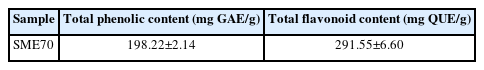
Total phenolic and flavonoid contents of Salvia microphylla ethanol extract
나. S. microphylla 70% 에탄올 추출물의 DPPH radical 소거능 평가
Free radical은 노화를 일으키는 원인 물질로 알려져 있으며, DPPH에 의한 free radical 소거능은 활성산소종(ROS)으로 항산화 물질의 항산화력을 확인하는데 널리 사용되고 있다(Choi & Moon, 2017). S. microphylla 70% 에탄올 추출물의 DPPH radical 소거활성은 50-400 μg/mL에서 농도의존적으로 증가하고 그 이상의 농도에서는 변화 없이 소거활성을 나타내었다. 200 μg/mL에서 91.76±0.05%, 400 μg/mL에서 92.99±0.14% 및 600 μg/mL에서 92.21±0.08%의 소거활성을 나타내었다(Figure 1).
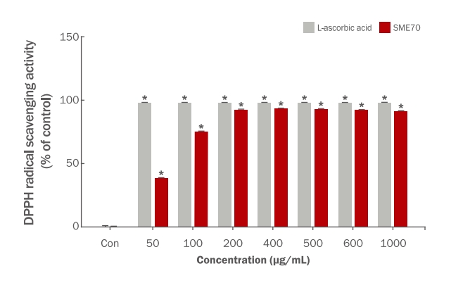
DPPH radical scavenging activity of Salvia microphylla ethanol extract.
DPPH radical scavenging assays were conducted to investigate the antioxidant effects of SME70 at various concentration. The values are then expressed as mean±SD of three independent experiments. *p<0.05 compared with untreated control. Con: none treated control; SME70, Salvia microphylla 70% ethanol extract.
총 폴리페놀 함량은 높을수록 항산화능이 우수하며, 플라보노이드는 구조에 의해 항산화 및 항균 등의 여러 생리활성이 있는 것으로 보고되었다(Bray & Thorpe, 1954; Middleton & Kandaswami, 1994; Kim & Hong, 2021). 따라서 총 폴리페놀 및 플라보노이드 함량에 상응하여 S. microphylla 70% 에탄올 추출물은 우수한 항산화능을 나태내어 화장품 및 식품에서 항산화제로서 도움을 줄 수 있을 것으로 사료된다.
다. S. microphylla 70% 에탄올 추출물의 ABTS radical 소거능 평가
ABTS radical은 비극성 물질에만 반응하는 DPPH radical과 달리 극성 및 비극성 물질에 모두 반응하며, DPPH radical과 반응하지 않는 비극성 물질이 ABTS radical과는 반응하여 소거되어진다고 알려져 있다(Kim et al., 2022). 또한 ABTS는 DPPH radical 소거능 측정법과 함께 항산화능을 측정하는데 널리 사용되는 방법 중 하나이다(Lee & Ahn, 2022). S. microphylla 70% 에탄올 추출물의 ABTS radical 소거활성은 50-600 μg/mL에서 농도의존적으로 하고 그 이상의 농도에서는 변화 없이 소거활성을 나타내었다. 200 μg/mL에서 77.37±2.69%, 400 μg/mL에서 98.89±0.18% 및 600 μg/mL에서 99.37±0.12%의 소거활성을 나타내었다(Figure 2).

ABTS radical scavenging activity of Salvia microphylla ethanol extract.
ABTS radical scavenging assays were conducted to investigate the antioxidant effects of SME70 at various concentration. The values are then expressed as mean±SD of three independent experiments. *p<0.05 compared with untreated control. Con: none treated control; SME70, Salvia microphylla 70% ethanol extract.
식물내에 존재하는 총 폴리페놀 및 플라보노이드 함량에 따라 항산화 활성과 생리활성이 증가한다고 보고되었다(Kim & Hong, 2021). 또한 DPPH와 ABTS 라디컬 소거능은 페놀성 물질 함량이 높을수록 증가됨으로 free radical 물질인 DPPH와 ABTS의 소거활성은 유의적인 상관관계를 갖는 것으로 알려져 있다(Chung & Kim, 2023; Lee et al., 2012). 따라서 총 폴리페놀 및 플라보노이드 함량에 상응하여 S. microphylla 70% 에탄올 추출물의 항산화능이 우수하다고 사료된다.
2. S. microphylla 70% 에탄올 추출물의 세포 생존율 확인
S. microphylla 70% 에탄올 추출물의 섬유아세포(CCD-986sk)의 세포 생존율을 측정하기 위하여 MTT assay를 이용하여 50-1,000 μg/mL로 처리하여 48 h 배양 후 대조군과 비교하였다(Figure 3). S. microphylla 70% 에탄올 추출물의 세포 생존율은 50 μg/mL에서 100.79±4.95%, 100 μg/mL에서 104.08±5.03% 및 200 μg/mL에서 99.56±2.77%을 나타내었으며, 400 μg/mL에서 78.97±2.20% 및 1000 μg/mL에서 40.26±1.55%로 세포 생존율이 감소되었다. 따라서 S. microphylla 70% 에탄올 추출물의 세포 생존율 결과를 바탕으로 50-200 μg/mL 농도범위에서 pro-collagen 합성과 MMP-1의 활성 억제 측정에 적용하였다.
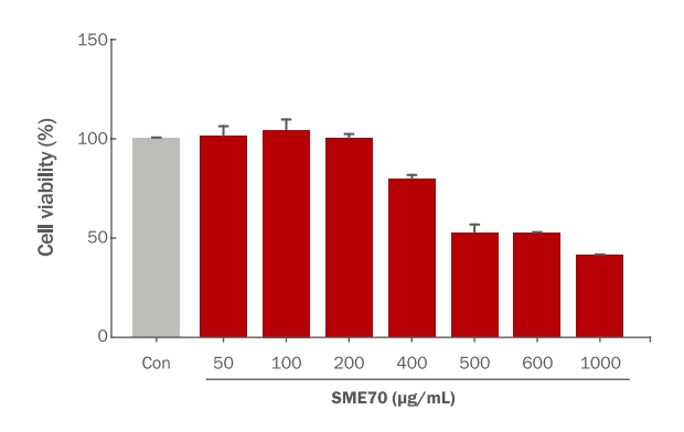
Effect of Salvia microphylla ethanol extract on cell viability in human fibroblasts.
CCD-986sk cells were treated with various concentration of SME70 (50–1000 μg/mL) using MTT assay. The responses in the control in each test are expressed as 100%. Each value expresses the mean±SD of three independent experiments. Con. none treated control; SME70, Salvia microphylla 70% ethanol extract.
3. S. microphylla 70% 에탄올 추출물의 pro-collagen type-Ⅰ 합성 확인
세포외기질(extracellular matrix, ECM)의 주요 구성 성분인 콜라겐(collagen)은 진피층의 섬유아세포에서 생성되는 주요 기질단백질로 피부의 주름생성과 탄력의 조절에 기여한다(Kim & Yoon, 2013). Collagen은 전구체인 procollagen으로부터 합성되어, 아미노 말단과 카복시 말단에 propeptide라는 peptide sequence를 가지고 있으므로 collagen 합성양은 propeptide 양의 측정을 통해 생합성 효과를 판단할 수 있다(Tsuji-Naito et al., 2010; Kim et al., 2019).
S. microphylla 70% 에탄올 추출물의 주름개선 효능에 관한 pro-collagen 합성은 농도의존적으로 50 μg/mL에서 110.92±0.99%, 100 μg/mL에서 120.94±4.66% 및 200 μg/mL에서 140.74±5.31%로 증가하는 것을 유의하게 나타내었다(Figure 4). 이와 같이 pro-collagen 합성 증가 결과는 S. microphylla 70% 에탄올 추출물이 섬유아세포에서 콜라겐 합성을 유도하는 능력을 가지고 있음을 시사한다.
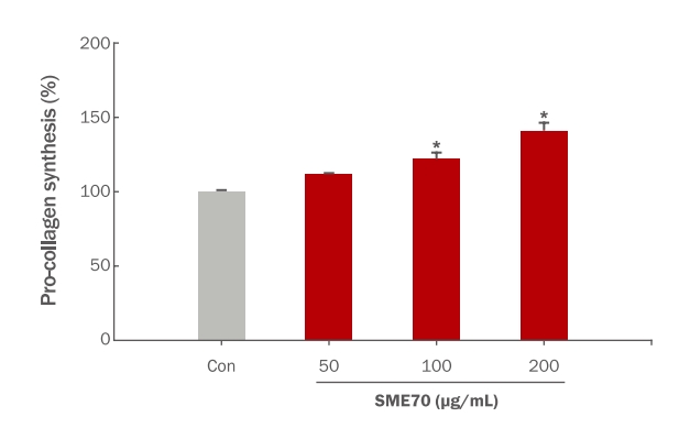
Effect of Salvia microphylla extract on pro-collagen synthesis in human fibroblasts.
Pro-collagen synthesis effect of SME70 on CCD-986sk cells. After CCD-986sk cells (1×10⁶ cells) were treated with 50, 100, and 200 μg/mL of extracted of SME70 for 48 h. Each value expresses the mean±SD of three independent experiments. *p<0.05 compared with untreated control. Con: none treated control; SME70: Salvia microphylla 70% ethanol extract.
5. S. microphylla 70% 에탄올 추출물의 MMP-1 활성 억제 효과
피부에서 TypeⅠ collagen의 합성과 그 분해효소인 MMP-1 활성이 균형을 이루고 있지만, 내외적인 원인으로 인해 TypeⅠ과 Type Ⅲ collagen 합성이 저하되고, MMP-1 활성이 증가되는 것으로 알려져 있다(Lee & Lee, 2013). S. microphylla 70% 에탄올 추출물에 의한 MMP-1 활성은 농도의존적으로 50 μg/mL에서 97.69±0.48%, 100 μg/mL에서 76.52±1.28% 및 200 μg/mL에서 65.36±1.40%로 감소하였다(Figure 5). 따라서 S. microphylla 70% 에탄올 추출물에 의해 MMP-1 활성이 최대 35% 억제하는 것을 확인하였다. 이와 같은 실험 결과에 의해 섬유아세포에서 pro-collagen 합성 증가는 S. microphylla 70% 에탄올 추출물의 MMP-1 활성 억제에 따른 collagen 합성량의 증가로 판단된다. 따라서 S. microphylla 70% 에탄올 추출물은 기능성 화장품 소재로서 피부탄력 및 주름개선에 기여할 것으로 사료된다.
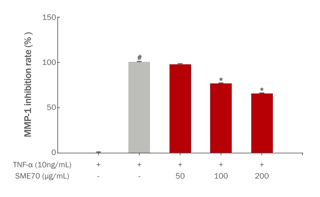
Effect of Salvia microphylla extract on MMP-1 inhibition rate in human fibroblasts (CCD-986sk).
After CCD-986sk cells (1×10⁶ cells) were incubated with or without SME70 (50–200 μg/mL) in the presence or absence of TNF-α (10 ng/mL) for 48 h. Each value expresses the mean±SD of three independent experiments. #p<0.05 compared with untreated control, *p<0.05 compared with TNF-α treated control. SME70, Salvia microphylla 70% ethanol extract; TNF-α: tumor necrosis factor-α.
Conclusion
본 연구에서는 S. microphylla 70% 에탄올 추출물을 이용하여 항산화, pro-collagen 합성 및 MMP-1 활성 억제 효과 등을 조사하여 피부탄력 및 주름개선 효과를 확인하고자 하였다. 폴리페놀과 플라보노이드 함량은 각각 198.22±2.14 mg GAE/g과 291.55±6.60 mg QUE/g의 함량을 나타내었다. DPPH radical 소거능은 최대 400 μg/mL 농도에서 92.99±0.14%의 소거활성을 확인하였으며, ABTS radical 소거능은 최대 600 μg/mL 농도에서 99.37±0.12%의 소거활성을 확인하였다. 총 폴리페놀 및 플라보노이드 함량에 따라 항산화 활성과 생리활성이 증가(Kim & Hong, 2021)하는 것과 같이 S. microphylla 70% 에탄올 추출물의 항산화 활성이 뛰어난 것으로 확인하였다. S. microphylla 70% 에탄올 추출물의 주름개선 효능을 확인하고자 pro-collagen 합성을 확인한 결과, pro-collagen 합성은 농도의존적으로 증가하여 최대 200 μg/mL에서 140.74±5.31%의 합성 증가를 확인하였다. Collagen 분해요소인 MMP-1 활성은 농도 의존적으로 감소하여 200 μg/mL에서 65.36±1.40%로 MMP-1 활성이 최대 35% 억제하는 것을 확인하였다. 이와 같은 실험 결과에 의해 섬유아세포에서 pro-collagen 합성 증가는 S. microphylla 70% 에탄올 추출물의 MMP-1 활성 억제에 따른 collagen 합성량의 증가로 판단된다. 따라서 본 연구에 사용된 S. microphylla 에탄올 추출물이 피부탄력 및 주름개선용 기능성화장품 소재로 응용이 가능할 것으로 판단된다.
Acknowledgements
이 성과는 정부(과학기술정보통신부)의 재원으로 한국연구재단의 지원을 받아 수행된 연구임(No. 2021R1G1A1094165).
Notes
Author's contribution
IHC designed all the experimental investigations. Biological activity experiments were conducted together by HMJ, and IHC oversaw the project and contributed to all aspects of analysis and experimental design.
Author details
Hyeon Mi Jo (Graduate Student), Department of Biomaterials Science, Gyeongsang National University, 33 Dongjin-ro, Jinju-si, Gyeongsangnam-do, 52725, Korea; In Ho Choi (Professor), Department of Plant and Biomaterials Science, Gyeongsang National University, 33 Dongjin-ro, Jinju-si, Gyeongsangnam-do, 52725, Korea.