효모 발효 식물 뿌리 혼합 추출물의 피부세포에 대한 생물학적 활성

요약

목적

혼합 식물 뿌리 추출물 및 발효 혼합 식물뿌리 추출물의 생물학적 활성을 확인하기 위하여 체외 실험을 통하여 항노화 및 항염증 효과를 확인하였다.

방법

DPPH 라디칼 소거 활성 평가를 통하여 항산화 활성이 우수한 식물 뿌리추출물을 선별하였으며, 선별된 식물 뿌리의 혼합 추출물 (MC) 및 S. cerevisia를 이용하여 식물 뿌리 혼합추출물의 발효물을 (MF) 제조하였다. 인체 피부 섬유아세포에서 MC 및 MF의 COL1A1HAS2 mRNA 발현에 대한 영향을 확인하였으며, 세포 증식 실험을 통하여 세포 이동능을 평가하였다. MC 및 MF의 항염증 효과를 확인하기 위하여 인체 각질 세포에서 RT-PCR을 통하여 COX2, TNF-α, IL6의 발현을 확인하였다. 실험 결과는 평균치와 표준편차로 나타내었고, 결과는 t-test를 사용하여 분석하였다.

결과

DPPH 라디칼 소거 활성 평가를 통하여 15종의 뿌리 추출물 중 5 종을 선별하였다. MC 및 MF는 COL1A1HAS2의 발현을 농도의존적으로 증가시켰으며, 세포 증식효과를 보여주었다. 또한 MC 및 MF는 염증 관련 유전자인 TNF-α, IL6, COX2의 발현을 농도 의존적으로 저해하였다. 모든 실험에서 MF는 MC와 비교하여 더 좋은 효과를 보여주었다.

결론

서양민들레뿌리줄기/뿌리추출물, 우엉뿌리추출물, 지모뿌리 추출물, 칡추출물 및 연꽃뿌리추출물의 혼합 발효물은 항노화 및 항염효과를 갖고 있어 기능성 화장품 원료로 적용 가능하다.

Abstract

Purpose

To verify the biological effects including anti-aging and anti-inflammatory effects, of fermented plant root extract mixtures that were measured in vitro.

Method

To select the most effective plant root extract, the effects of DPPH free radical scavenging were measured. The mixture (MC) of selected plant roots was fermented with Saccharomyces cerevisiae (S. cerevisiae) (MF). Reverse transcription (RT)-PCR was performed on human dermal fibroblast to measure the effects of MC and MF on the mRNA expression of COL1A1 and hyaluronic acid synthase 2 (HAS2) and on the anti-inflammatory-related gene expressions such as cyclooxygenase-2 (COX-2), interleukin-6 (IL-6), and tumor-necrosis factor (TNF)-α. Additionally, we also tested the cell renewal and proliferation for skin cell migration. Student's t-test was performed for statistical analysis, and results were expressed as mean ± standard deviation.

Results

Among 15 root extracts, those of Taraxacum officinale (T. officinale) rhizome, Arctium lappa (A. lappa), Anemarrhena asphodeloides (A. asphodeloides), Pueraria lobata (P. lobata), and Nelumbo nucifera (N. nucifera) were selected based on the effect of DPPH free radical scavenging. The mRNA expression levels of COL1A1 and HAS2 were increased by MC and MF in a concentration-dependent manner. MC and MF showed cell renewal and proliferation. Inflammation-related genes were inhibited by MC or MF in a concentration-dependent manner (TNF-α, IL-6, and COX-2). MF showed significantly better efficacy than MC.

Conclusion

Mixed root extracts of T. officinale rhizome/root, A. lappa, A. asphodeloides, P. lobata, and N. nucifera fermented with S. cerevisiae enable cell renewal, have anti-aging and anti-inflammatory effects, and can be used as an active cosmetic raw materials.

中文摘要

目的

为了确认混合植物根提取物和发酵混合植物根提取物的生物活性,通过体外实验证实了抗衰老和抗炎作用。

方法

通过对DPPH自由基清除活性的评价,筛选出具有优异抗氧化活性的植物根提取物,并利用所选择的植物根混合提取物(MC)和酿酒酵母制备了混合植物根提取物(MF)的发酵产物。确认MC和MF对人皮肤成纤维细胞COL1A1HAS2 mRNA表达的影响,并通过细胞增殖实验评价了细胞迁移能力。为了证实 MC和MF的抗炎作用,通过RT-PCR证实了 COX2TNF-α、IL6在人角质形成细胞中的表达。实验结果用平均值和标准差表示,结果采用t检验分析。

结果

通过对DPPH自由基清除活性的评价,选择了15种根提取物中的5种。 MC和MF以浓度依赖性方式增加COL1A1HAS2的表达,并表现出细胞增殖作用。此外,MC和MF以剂量依赖性方式抑制炎症相关基因TNF-α、IL6、COX2的表达。在所有实验中,与MC相比,MF显示出更好的效果。

结论

蒲公英根茎/根提取物、牛蒡根提取物、知母根提取物、野葛根提取物和莲根提取物的混合发酵产物具有抗衰老和抗炎作用,可作为功能性化妆品原料应用。

Introduction

Skin aging shows structural, and functional changes that are either intrinsic or extrinsic. Intrinsic aging is defined as the decreased skin structure and physiological activity over time, while extrinsic aging, or photoaging, is caused by external stresses such as ultraviolet and reactive oxygens (Helfrich et al., 2008).
The keratinocytes from the basal layer of the skin move toward the surface of the stratum corneum. These are removed through desquamation, as the skin regenerates over time, a period called skin turnover. The life span of keratinocytes in the human skin is approximately 20-28 days (Ho & Dreesen, 2021). Skin renewal by the keratinocytes slows down as the skin becomes older (Choi et al., 2017). Lifestyle habits such as insufficient sleep, alcohol drinking, and smoking retard of skin cell regeneration. Skin-related problems such as wrinkles and freckles occur due to the delay of skin turnover.
Studies on herbs and its benefit for skincare have recently increased, particularly due to the interest of cosmetic consumers in natural products (Shim, 2021; Zhang et al., 2018; Moon et al., 2017; Di Napoli & Zucchetti, 2021; Sivamaruthi et al., 2018). The benefits of mixed plant extracts and the mixed extracts have been reported to be better than those of individual extracts (Choi, 2022; Kim et al., 2019; Lee et al., 2021).
By fermentation, certain common substrate components are degraded or transformed into compatible components. This process can generally increase the physiological and biochemical activities of biological substrates by modifying naturally occurring molecules and can be used to develop new pharmaceutical and cosmetic formulations (Kim, 2011; Chiba, 2007; Um et al., 2017; Hussain et al., 2016). The cytotoxicity of certain herbal extracts can be reduced or eliminated after treatment using various microbial species. Some probiotics, such as Bifidobacterium bifidum and Saccharomyces cerevisiae, can produce new components by fermentation or reduce the cytotoxicity of herbal extracts (Shim et al., 2019; Hussain et al., 2016; Wang et al., 2016).
In the present study, the effects of plant roots extracts of Taraxacum officinale, Arctium lappa, Anemrrhena asphodeloides, Pueraria lobata, and Nelumbo nucifera fermented with S. cerevisiae on the expression of anti-aging and inflammatory-related genes were evaluated using human keratinocytes and fibroblasts to evaluate their potential as a cosmetic raw material.

Materials and Method

1. Plant roots and extraction

Each plant root was acquired from Shenzhen Mannay Cosmetics Co. Ltd., and was dried and ground before extraction (Table 1). The resulting powder was extracted in 20 volumes of 70% alcohol at room temperature for 3 h and filtered using a filter paper (No. 2 qualitative filter papers, Whatman, England). The resulting filtrates were concentrated under a vacuum using a rotary evaporator (N-1110; EYELA, USA) until alcohol was eliminated. The concentrated extracts were stored at -20°Cuntil further use.
The select root powders were mixed at equal amounts and extracted as described above. To prepare the fermented root mixture extracts, pre-cultured S. cerevisiae (1×10⁶/mL) was inoculated on the root mixture extracts and fermented at 28°C with 30 rpm for 3 days. To terminate the fermentation, the cultures were boiled at 100°C for 30 min and filtered using a 0.45 μm filter.
The international nomenclature cosmetic ingredient (INCI) of each plant root extract used in this study is described in Table 1.

2. DPPH free radical-scavenging activity assay

The free radical-scavenging activity of plant root extracts was determined using a 2,2-diphenyl-1-picrylhydrazyl (DPPH; Sigma Aldrich, USA) assay. 50 µL of the diluted extracts were mixed with 100 µL of 0.1 mM DPPH solution. The DPPH solution without the test sample was used as a control. The mixture was incubated for 30 min at room temperature and subsequently measured for its absorbance at 515 nm. The antioxidative activity was calculated using the formula below and expressed as the percentage of DPPH radical elimination:
[(Ablank-Asample)/Ablank]×100 (%)
where Ablank is the absorbance of the blank DPPH solution and Asample is the absorbance of the DPPH solution after the addition of test sample.

3. Cell lines

Human dermal fibroblasts (HDF; Thermo Fisher Scientific, USA) were cultured in Fibroblast Basal Medium (FBM 106; Gibco, USA) supplemented with low serum growth supplement (LSGS; Gibco), 100 IU/mL penicillin, and 100 μg/mL streptomycin. Human epidermal keratinocytes (HEKa; Invitrogen, USA) were cultured in Dulbecco's modified Eagle's medium (DMEM; Gibco, Thermo Fisher Scientific, USA) supplemented with antibiotics (100 IU/mL penicillin and 100 μg/mL streptomycin) and 10% fetal bovine serum (FBS; Gibco BRL, USA). The cells were maintained at 5% CO2 at 37°C. Confluent monolayer cultures of HDF and HEKa were trypsinized with 0.25% trypsin-EDTA.

4. Cell viability

Cell viability was determined by measuring the conversion of 3-(4,5-dimethyl-2-thiazolyl)-2,5-diphenyl-2H-tetrazolium bromide (MTT) into formazan via mitochondrial oxidation according to a modified Mosmann's method (Mosmann, 1983). HEKa and HDF were seeded at 1×10⁵ cells/well in a 96-well plate and cultured for 18 h. The prepared sample was added in 0, 1, 2, and 5% concentrations. After incubating for 24 h, the medium was gently removed, the cells were washed twice in phosphate-buffered saline (PBS), and then the wells were filled with 5 mg/mL of solution MTT (Sigma Aldrich) as the new medium. Afterward, formazan crystals produced from MTT were solubilized in 150 μL dimethyl sulfoxide (DMSO; Sigma Aldrich). The absorbance of each well was then measured at 570 nm using a microplate reader (Epoch2C, USA). The optical density of formazan formed in control cells without sample was considered as 100% viability.

5. Wound-healing (cell renewal) test

To examine cell renewal, HEKa was subjected to a wound-healing assay. The cells were inserted on a 12-well plate and then incubated for 4 h under cell culture conditions. On the removal of the culture medium, a "wound gap" in a cell monolayer was created by scratching, and then 2% of test samples along with a new culture medium were added to the cells; the plate was incubated again for 18 h. Four hours after incubation, cell migration was analyzed to measure the cell renewal effect.

6. Cell proliferation

To examine the cell proliferation of the test sample, CytoSelectTM BrdU Cell Proliferation ELISA Kit (Cell Biolabs Inc., USA) was used. The cells were inserted on a 96-well plate and then incubated for 18 h under cell culture conditions. Upon removal of the culture medium, the cells were washed with PBS, and then test samples along with a new culture medium were added to the cells; the plate was again incubated for 24 h. Afterward, a BrdU Solution was added to the wells, and the plate was again incubated at 37°C and 5% CO2 in a humidified incubator for 4 h. In each tested well, 100 µL of diluted anti-BrdU antibody and secondary antibody HRP conjugate were added. After incubation for 30 min, absorbance was measured according to the manufacturer's instructions. The cell proliferation rate of each sample treated was calculated based on the blank.

7. RNA isolation and revers transcription (RT)-PCR

To determine the mRNA expression of COL1A1, hyaluronic acid synthase2 (HAS2), tumor-necrosis factor-α (TNF-α), interleukin-6 (IL-6), and cyclooxygenase-2 (COX-2), cells were cultured in Medium 106 supplemented with 1×LSGS, 100 IU/mL penicillin, and 100 μg/mL streptomycin at 5% CO2 at 37°C for 24 h. The medium was gently removed, and the cells were treated with 0.1%, 0.5%, 1%, and 2% concentrations of the sample and then cultured for 48 h. Total RNAs from each well were isolated using the TransZol reagent and obtained to generate the cDNA using the PrimeScript 1st cDNA synthesis kit, as recommended by the manufacturer's instructions. Amplifications were performed in a thermal cycler. The experiment consisted of 35 cycles: denaturation at 95°C for 1 min, annealing at 50-60°C for 30 s, and extension at 72°C for 1 min. The sequences of primer pairs, and further details of PCR, are given in Table 2. Glyceraldehyde 3-phosphate dehydrogenase (GAPDH) was used as a control gene. PCR products were electrophoresed on a 1% agarose gel and stained with ethidium bromide to analyze the relative ratio of changes in the target gene in comparison to those of the control.

8. Statistical analysis

The results obtained were expressed as mean±standard deviation (SD) from at least three independent experiments. The student's t-test with Microsoft Excel (Microsoft, USA) was used to analyze the data. p<0.05 was considered to indicate statistical significance.

Results and discussion

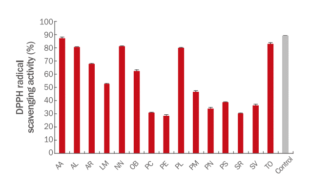
In the cosmetic industry, natural ingredients have been increasingly a point of concern. In this study, we focused on plant roots due to their medicinal use in Asian cultures (Bark et al., 2010). We prepared 15 plant root extracts (Table 1) and evaluated the antioxidant effect using DPPH free radical scavenging assay. The tested plant root extracts show an antioxidant effect at 10% treatment (Figure 1). Among them, only five show more than 80% of DPPH free radical-scavenging activity: Taraxacum offininale, Arctium lappa, Anemrrhena asphodeloides, Pueraria lobata, and Nelumbo nucifera.
The synergic effect of a mixture of plant extract and the fermented mixture of plant extract have been reported (Kim et al., 2019; Um et al., 2017). Five roots were selected to be mixed and extracted (MC). In addition, MC was fermented using S. cerevisiae (MF). Fermentation is a strategy to improve efficacy and reduced toxicity product and is frequently used to manufacture cosmetic raw materials.
The results showed that MC and MF treated on HDF and HEKa had more than 80% of cell viability at 2% concentration and following works were carried out in this range (Figure 2).
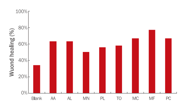
The effects of each root extract, MC, and MF on cell renewal were measured in HEKa cells: 34-63% by each root extract and 67% of cell renewal activity by MC-treated HEKa cells, which is similar to that by positive controls. MF-treated HEKa cells recovered the wound gap by approximately 77%, which was approximately 20% higher than that of MC. Thus, MF promotes skin cell regeneration and activates the proliferation of the skin cells (Figure 3).
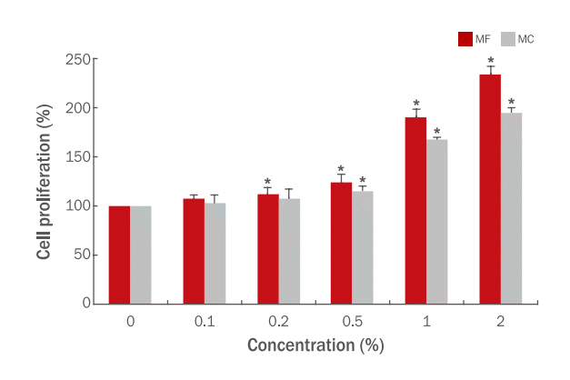
Our results also confirmed that MC and MF affect cell proliferation in HEKa cells. Figure 4 shows that MC and MF have cell renewal and cell proliferation in a dose-dependent manner. Results indicated that cell proliferation was significantly increased by MF-treated cells compared to that by MC-treated cells (p<0.05). Cell proliferation in HDF cells treated with 2% MF and 2% MC was significantly higher than that in untreated cells (234.06% and 195.38%, respectively); MF treatment results in approximately 38.68% higher than that of MC treatment at 2% treatments. In terms of efficacy, our result was similar with that of Hsu and Chiang when they compared the effect of fermented Radix astragali with Bacillus subtilis natto and non-fermented R. astragali, which is well-known for its medicinal effect. The fermented R. astragali has a better anti-aging effect despite the reduction in concentration of main isoflavonoids such as galycosin, formononetin, and ononin (Hsu & Chiang, 2009).
Collagen is composed of three polypeptide α-chains that form a triple helical structure. COL1A1 is a gene-encoding type I collagen that is one of fibril-forming collagen (Yue, 2014; Ricard-Blum, 2011). The effects of MC and MF on COL1A1 mRNA expression were evaluated in HDF cells using RT-PCR. The mRNA expression levels of COL1A1 in MC- and MFtreated HDF cells increased in a dose-dependent manner (Figure 2). Similar to the cell proliferation test result, the effect of MF on COL1A1 mRNA expression was higher than that of MC with a significantly different (p<0.05). The mRNA expression levels of COL1A1 in HDF cells treated with 2% MC and MF were 157.37% and 195.11%, respectively, while that of a positive control (50 μM retinyl palmitate treatment) was 220.57% (Figure 5A). COL1A1 mRNA expression in MF-treated cells was approximately 24% higher than that of MC-treated cells at 2% (p<0.05).
In the skin, gene expression of HAS2 is stimulated by keratinocyte growth factor, which activates keratinocyte migration and stimulates wound healing (Papakonstantinou et al., 2012). We measured the effect of MC and MF on HAS2 mRNA expression. HAS2 mRNA expression was increased in a dose-dependent manner, with MF-treated cells having significantly higher results than those of MC-treated cells (p<0.05). HAS2 expression levels of HDF cells treated with 2% MC and MF were 221.71% and 256.64%, respectively, and that of 50 μg/mL hyaluronic acid was 267.39% (Figure 5B).
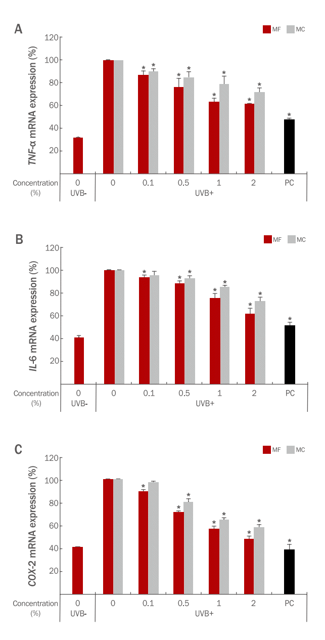
Inflammation of the skin was caused by TNF-α, ILs, and other cytokines that could be upregulated by environmental or physiological stresses, thereby restricting the growth of skin cells while inhibiting skin inflammation and aging. Inflammation is one of the intrinsic reasons for aging, and to maintain a healthy skin, inflammation control is important (Baylis et al., 2013). The effect of MC and MF on inflammation-related gene expressions such as TNF-α, IL6, and COX2 was measured in HEKa cells (Figure 6). TNF-α mRNA expression in HDF cells after treatment of 2% of MC was measured as 157.37% of UV-irradiated control, whereas the same concentration of MF treatment was measured as 195.11%. A positive control treatment with 50 μM retinyl palmitate resulted in a TNF-α expression level of 220.57% (Figure 6A).
At 2% treatment of MC, IL6 mRNA expression in HDF cells was measured as 157.37% of UVB-irradiated control, whereas the same concentration of MF treatment was measured as 195.11%. A positive control treatment with 50 μM retinyl palmitate resulted in an IL6 expression level of 220.57% (Figure 6B).
The effect of MC and MF on COX2 mRNA expression in HDF cells also similar with other inflammatory-related biomarkers (157.37% and 195.11%, respectively, of UVB-irradiated control). A positive control treatment with 50 μM retinyl palmitate resulted in a COX2 expression level of 220.57% (Figure 6C). The expression of TNF-α, IL6, and COX2 mRNA in MC- or MFtreated HEKa cells was inhibited in a concentration-dependent manner.
NN root contains betulinic acid, rutin, isoquercetin, and other flavonoids that have antioxidant, anti-inflammatory, and cytoprotective activities (Sruthi et al., 2019). Petkova et al. reported the phytochemical composition of AL root extracts and their antioxidant effects. AL roots contain phenolic compounds such as chlorogenic acid, caffeic acid, and p-coumaric acid (Petkova et al., 2022), and these compounds are well-known antioxidants (Predes et al., 2011; Rosalia et al., 2010). Phytochemicals such as carotenoids, flavonoids, and phenolic compounds have a number of medicinal properties such as antioxidant, antiviral, antifungal, and antibacterial effects (Di Napoli & Zucchetti, 2021). Wang et al. isolated saponins from AA roots and investigated the isolated compounds that inhibit LPS-induced NO production (Wang et al., 2018). PL root, known as Kuzu, has been used as a traditional medicine for treating cardiovascular diseases and type 2 diabetes mellitus in China (Luo et al., 2007). PL root contains antioxidants, isoflavones, triterpene, and their glycosides and have anti-inflammatory and anti-apoptosis properties (Zhang et al., 2018; Wong et al., 2011). Each root contains various active compounds that possess biological activities.
In this study, we prepared a mixture of extracts using five roots selected from 15 roots based on DPPH radical scavenging activity, and the effect of MC and MF were compared on the mRNA expression of COLA1A1 and HAS2 and cell proliferation, as well as their effect of MC and MF on the expression of inflammation-related mRNA such as TNF-α, IL-6, and COX2. Based on our results, the treatment of MC and MF could enhance anti-aging properties by promoting COL1A1 and HAS2 mRNA expression and inducing a positive effect on cell migration. Fermentation of the mixture of the selected roots extracts significantly contributed to the increased anti-aging capabilities.

Conclusion

Of 15 plant root extracts, only five root extracts were selected based on the result of DPPH free radical scavenging activity: T. officinale, A. lappa, A. asphodeloides, P. lobata, and N. nucifera. The mixture of selected root extracts (MC) and their fermented products (MF) with S. cerevisiae improved COL1A1 and HAS2 mRNA expressions and cell proliferation efficacy in HDF. MC and MF also showed anti-inflammatory effects in human keratinocytes and inhibited the mRNA expression of TNF-α, IL6, and COX2. Furthermore, MF had better efficacies for antiaging and anti-inflammation than MC. Based on these results, MF has better efficacy than MC and can be used as an active material for cosmetics with anti-aging and anti-inflammation effects.

Acknowledgements

This study was supported by Shenzhen Mannay Cosmetic Co., Ltd (Shenzhen, China).

NOTES

Author's contribution
HL, YJK designed the study. HL, YJK carried out the extraction and biochemical assays. HL and WHC oversaw the project. HL wrote the manuscript with assistance from YJK. All authors read and approved the final manuscript.
Author details
Hyunsang Lee (R&D Director), Mannay Asia R&D Center, 16 Heungandae-ro, 427 Beon-gil, Dongan-gu, Anyang-si, Gyeonggi-do 14059, Korea; Yun Jeong Kim (Principal Researcher), Mannay Asia R&D Center, 16 Heungandae-ro, 427 Beon-gil, Dongan-gu, Anyang-si, Gyeonggi-do 14059, Korea; Wai Ho Cheung (Executive Director), Shenzhen Mannay Cosmetic Co., Ltd, #A, Jiahe Huaqiang Building, Shennan Middle Road, Futian District, Shenzhen, Guangdong 518000, China.

Figure 1.

DPPH radical scavenging activity of plant root extracts.

DPPH free radical-scavenging activity of plant root extracts (10%). Quercetin (1000 μg/mL) was used as a positive control The results are expressed as the mean±SD of three independent experiments. AA, Anemarrhena asphodeloides root extract; AL, Arctium lappa root extract; AR, Althaea rosea root extract; LM, Lepidium meyenii root extract; NN, Nelumbo nucifera root extract; OB, Oenothera biennis root extract; PC, Polygonum cuspidatum root extract; PE, Pachyrrhizus erosus root extract; PL, Pueraria lobata root extract; PM, Polygonum multiflorum root extract; PN, Panax notoginseng root extract; PS, Paeonia suffruticosa root extract; SR, Sedum rosea root extract; SV, Sciadopitys verticillata root extract; TO, Taraxacum officinale rhizome/root extract.
ajbc-20-3-349f1.tif
Figure 2.

Cell viability of root extracts treated cells.

Cells are treated with each root extract 1, 2, and 5% concentrations for 24 h. Cytotoxicity was measured using the MTT assay. The results are expressed as the mean±SD of three independent experiments. A, HDF cells; B, HEKa cells. *Statistically significant (p<0.05).
ajbc-20-3-349f2.tif
Figure 3.

Cell regeneration effect of plants roots extract, MC, and MF in HEKa cells.

“Wounds gap” in a cell monolayer is created by scratching and test samples. Cells are treated with 2% of each sample for 18 h. AA, Anemarrhena asphodeloides root extract; AL, Arctium lappa root extract; NN, Nelumbo nucifera root extract; PL, Pueraria lobata root extract; TO, Taraxacum officinale rhizome/root extract; MC, mixed plant root extract; MF, fermented mixed roots extract. 0.1% of madecassoside is used for a positive control (PC).
ajbc-20-3-349f3.tif
Figure 4.

Effect of MC and MF on cell proliferation in HEKa cells.

Various concentration of MC and MF (0.1, 0.5, 1, and 2 %) were treated on HEKa cells and measured the cell proliferation activity using CytoSelect Brd U Cell proliferation ELISA Kit. Data are expressed as mean±SD of three independent experiments. *Statistically significant (p<0.05).
ajbc-20-3-349f4.tif
Figure 5.

Effect of MC and MF on COL1A1 and HAS-2 mRNA expression in HDF cells.

The expression levels of COL1A1 and HAS-2 were measured by RT-PCR in HDF treated with indicated increasing concentrations of MC or MF. Each value represents the mean±SD of three individual experiments. Retinyl palmitate (50 μM) was used as a positive control (PC1) for COL1A1. Hyaluronic acid (50 μg/mL) was used as a positive control (PC2) for HAS-2. *Statistically significant (p<0.05).
ajbc-20-3-349f5.tif
Figure 6.

Effect of MC and MF on inflammatory-related mRNA expression in HEKa cells.

The expression levels of TNF-α (A) IL6 (B) COX-2 (C) were measured by RT-PCR in HEKa treated with indicated increasing concentrations of MC or MF. Each value represents the mean±SD of three individual experiments. Allantoin (0.01 μg/mL) was used as a positive control (PC).
ajbc-20-3-349f6.tif
Table 1.
INCI of plant root extracts used in this study.
INCI Korean name Chinese name Abbreviations
Althaea rosea root extract 접시꽃뿌리추출물 蜀葵花提取物 AR
Anemarrhena asphodeloides root extract 지모뿌리추출물 知母根提取物 AA
Arctium lappa root extract 우엉뿌리추출물 牛蒡根提取物 AL
Lepidium meyenii root extract 마카뿌리추출물 迈因葶苈根提取物 LM
Nelumbo nucifera root extract 연꽃뿌리추출물 莲根提取物 NN
Oenothera biennis (Evening Primrose) root extract 달맞이꽃뿌리추출물 月见草根提取物 OB
Pachyrhizus erosus root extract 얌빈뿌리추출물 豆薯根提取物 PE
Paeonia suffruticosa root extract 모란뿌리추출물 牡丹根提取物 PS
Panax notoginseng root extract 삼칠뿌리추출물 三七根提取物 PN
Polygonum cuspidatum root extract 호장근뿌리추출물 虎杖根提取物 PC
Polygonum multiflorum root extract 하수오뿌리추출물 何首乌根提取物 PM
Pueraria lobata root extract 칡뿌리추출 물 野葛根提取物 PL
Sciadopitys verticillata root extract 금송뿌리추출물 金松根提取物 SV
Sedum rosea root extract 돌꽃뿌리추출물 玫瑰红景天根提取物 SR
Taraxacum officinale rhizome/root extract 서양민들레뿌리줄기/뿌리추출물 欧蒲公英根茎/根提取物 TO
Table 2.
Primer sequences
Primer Forward sequence Reverse sequences
GAPDH*1 ATT GTT GCC ATC AAT GAC CC AGT AGA GGC AGG GAT GAT GT
GAPDH*2 CTG GCA CCC AGC ACA ATG AAG ACC GAC TGC TGT CAC CTT CA
HAS2 GCT ACC AGT TTA TCC AAA CG GTG ACT CAT CTG TCT CAC CG
COL1A1 GGC CCA GAA GAA CTG GTA CA CGC TGT TCT TGC AGT GGT AG
IL6 ATG AAC TCC TTC TCC ACA AGC GC GAA GAG CCC TCA GGC TGG ACT G
COX2 GAA GAG CCC TCA GGC TGG ACT GT AGA TCA TCT CTG CCT GAG TAT CTT
TNF-α CAT TCT GGG AGG GGT CTT CC GGT TGA GGG TGT CTG AAG GA
AQP3 TGC AAT CTG GCA CTT CGC GCC AGC ACA CAC ACG ATA A

*1 for HDF;

*2 for HEKa;

GAPDH, glyceraldehyde-3-phosphate dehydrogenase; HAS 2, hyaluronic acid synthase 2; COL1A1, collagen type 1 alpha 1 chain; IL 6, interleukin 6; COX2, cyclooxygenase 2; TNF-α, tumor-necrosis factor-α.

References

Bark KM, Heo EP, Han KD, Kim MB, Lee ST, Gil EM, Kim TH. Evaluation of the phototoxic potential of plants used in oriental medicine. Journal of Ethnopharmacology 127: 11-18. 2010.
crossref pmid
Baylis D, Vartlett DB, Patel HP, Roberts HC. Understanding how we age: insights into inflammaging. Longevity and Healthspan 2: 8. 2013.
crossref pmid pmc
Chiba K. Development of functional cosmetic ingredients using lactic acid bacteria in Japan. Japanese Journal of Lactic Acid Bacteria 18: 105-112. 2007.
crossref
Choi JH, Song YS, Song K, Lee HJ, Hong JW, Kim v. Skin renewal activity of non-thermal plasma through the activation of β-catenin in keratinocytes. Scientific Reports 7: 6146. 2017.
crossref pmid pmc
Choi MJ. Prunus persica L., Nelumbo nucifera, Hibiscus mutabilis L., Agastanche rugosa, Wolfiporia extensa extracts to improve skin wrinkles. Asian Journal of Beauty and Cosmetology 20: 11-19. 2022.

Di Napoli A, Zucchetti P. A comprehensive review of the benefits of Taraxacum officinale on human health. Bulletin of the National Research Centre 45: 110. 2021.

Ferracane R, Graziani G, Gallo M, Fogliano V, Ritieni A. Metabolic profile of the bioactive compounds of burdock (Arctium lappa) seeds, roots and leaves. Journal of Pharmaceutical and Biomedical Analysis 51: 399-404. 2010.
crossref pmid
Helfrich YR, Sachs DL, Voorhees JJ. Overview of skin aging and photoaging. Dermatology Nursing 20: 177-183. 2008.
pmid
Ho CY, Dreesen O. Faces of cellular senescence in skin aging. Mechanisms of Ageing and Development 198: 111525. 2021.
crossref pmid
Hsu MF, Chiang BH. Effect of Bacillus subtilis natto-fermented Radix astragali on collagen production in human skin fibroblasts. Process Biochemistry 44: 83-90. 2009.
crossref
Hussain A, Bose S, Wang JH, Yadav MK, Mahajan GB, Kim H. Fermentation, a feasible strategy for enhancing bioactivity of herbal medicines. Food Research International 81: 1-16. 2016.
crossref
Kim CH. Development of cosmetic raw material using fermentation and bio-conversion technology. News & Information for Chemical Engineers 29: 42-48. 2011.

Kim HW, Kim DS, Sung NY, Han IJ, Lee BS, Park SY, Eom J, Suh JY, Park J, Yu AR, et al. Development of functional cosmetic material using a combination of Hippophae rhamnoides fruit, Rubus fruticosus leaf and Perillae folium leaf extracts. Asian Journal of Beauty and Cosmetology 17: 477-488. 2019.
crossref
Lee S, An H, Kim W, Lu X, Jeon H, Park HW, Ha J, Cho J. Antiaging effect of mixed extract from medicinal herbs. Asian Journal of Beauty and Cosmetology 19: 679-692. 2021.
crossref
Luo Q, Hao J, Yang Y, Yi H. Herbalogical textual research on “Gegen”. China Journal of Chinese Materia Medica 32: 1141-1144. 2007.
pmid
Moon JS, Lee JH, Kim YB. Study of antioxidation activity and melanocyte effect of Pueraria lobata root extract. Journal of the Korean Applied Science and Technology 34: 418-425. 2017.

Mosmann T. Rapid colorimetric assay for cellular growth and survival: application to proliferation and cytotoxicity assays. Journal of Immunological Methods 65: 55-63. 1983.
crossref pmid
Papakonstantinou E, Roth M, Karakiulakis G. Hyaluronic acid: a key molecule in skin aging. Dermato-Endocrinology 4: 253-258. 2012.
crossref pmid pmc
Petkova N, Hambarlyiska I, Tumbarski Y, Vrancheva R, Raeva M, Ivanov I. Phytochemical composition and antimicrobial properties of burdock (Arctium lappa L.) roots extracts. Biointerface Research in Applied Chemistry 12: 2826-2842. 2022.
crossref
Predes FS, Ruiz AL, Carvalho JE, Foglio MA, Dolder H. Antioxidative and in vitro antiproliferative activity of Arctium lappa root extracts. BMC Complementary and Alternative Medicine 11: 25. 2011.
crossref pmid pmc
Ricard-Blum S. v. Cold Spring Harbor Perspectives in Biology 3: a004978. 2011.
crossref pmid pmc
Shim SY, Park WS, Shin HS, OK M, Cho YS, Ahn HY. Physicochemical properties and biological activities of Angelica gigas fermented by Saccharomyces cerevisiae. Journal of Life Science 29: 1136-1143. 2019.

Shim JH. Anti-aging effect of ganoderol A in UVA-irradiated normal human epidermal keratinocytes. Asian Journal of Beauty and Cosmetology 19: 57-64. 2021.
crossref
Sivamaruthi BS, Chaiyasut C, Kesika P. Cosmeceutical importance of fermented plant extracts: a short review. International Journal of Applied Pharmaceutics 10: 31-34. 2018.
crossref
Sruthi A, Seeja TP, Aneena ER, Berin P, Deepu M. Insights into the composition of lotus rhizome. Journal of Pharmacognosy and Phytochemistry 8: 3550-3555. 2019.

Um JN, Min JW, Joo KS, Kang HC. Antioxidant, anti-wrinkle activity and whitening effect of fermented mixture extracts of Angelica gigas, Paeonia lactiflora, Rehmannia chinensis and Cnidium officinale. Korean Journal of Medicinal Crop Science 25: 152-159. 2017.
crossref
Wang GH, Chen CY, Lin CP, Huang CL, Lin CH, Cheng CY, Chung YC. Tyrosinase inhibitory and antioxidant activities of three Bifidobacterium bifidum-fermented herb extracts. Industrial Crops and Products 89: 376-382. 2016.
crossref
Wang Z, Cai J, Fu Q, Cheng L, Wu L, Zhang W, Zhang Y, Jin Y, Zhang C. Anti-inflammatory activities of compounds isolated from the rhizome of Anemarrhena asphodelioides. Molecules 23: 2631. 2018.
crossref pmid pmc
Wong KH, Li GQ, Li KM, Razmovski-Naumovski V, Chan K. Kudzu root: traditional uses and potential medicinal benefits in diabetes and cardiovascular disease. Journal of Ethnopharmacology 134: 584-607. 2011.
crossref pmid
Yue B. Biology of the extracellular matrix: an overview. Journal of Glaucoma 23: 520-523. 2014.

Zhang Z, Oh DS, Lee MS. The influence of Pueraria Lobata root extract upon anti-oxidant activity. Journal of The Korean Society of Cosmetology 24: 1098-1105. 2018.